

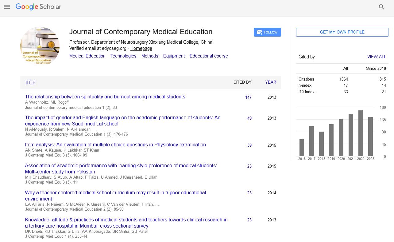


Perspective - Journal of Contemporary Medical Education (2022)
Pupil of the Eye and its Function, Size and Testing
Stepen Courn*Stepen Courn, Department of Chemistry, Indiana University-Purdue University, Indiana, USA, Email: stepenco@gmail.com
Received: 01-Sep-2022, Manuscript No. JCMEDU-22-74143; Editor assigned: 05-Sep-2022, Pre QC No. JCMEDU-22-74143 (PQ); Reviewed: 19-Sep-2022, QC No. JCMEDU-22-74143; Revised: 26-Sep-2022, Manuscript No. JCMEDU-22-74143 (R); Published: 03-Oct-2022
Description
The pupil of the eye is the portal that allows and regulates the flow of light to the retina. It is part of the process that allows us to perceive images. The pupil opens and closes to control the amount of light that is allowed to enter the eye.
The pupil is a black hole located in the center of the iris of the eye that allows light to enter the retina. It appears black because the light rays that enter the pupil are either absorbed by the tissues inside the eye directly or after diffuse reflections in the eye that mostly do not exit the narrow pupil. The term “pupil” was introduced by Gerard of Cremona.
In humans, the pupil is round, but its shape varies in different species; some cats, reptiles, and foxes have vertical slit pupils, goats have horizontal orientation, and some catfish have ring types. In optical terms, the anatomical pupil is the opening of the eye and the iris is the diaphragm. The outside pupil image is the entrance pupil, which does not exactly match the location and size of the physical pupil because it is magnified by the cornea. On the inner edge lies a prominent structure, the colorette, which marks the junction of the embryonic pupillary membrane that covers the embryonic pupil.
Pupil function
Together, the iris and pupil control how much light enters the eye. Using the camera analogy, the pupil is the opening of the eye and the iris is the diaphragm that controls the size of the aperture.
The size of the pupil is controlled by the iris muscles - one muscle narrows the pupil opening (makes it smaller) and the other iris muscle dilates the pupil (makes it bigger). This dynamic process of the iris muscles controls the amount of light that enters the eye through the pupil.
In low light conditions, the pupil dilates to allow more light to reach the retina and improve night vision. In bright light, the pupil constricts to limit the amount of light entering the eye (too much light can cause glare and discomfort, and can even damage the lens and retina).
Pupil size
Pupil size varies from person to person. Some have large pupils, and some have small pupils. Also, pupil size changes with age - children and young adults tend to have large pupils, while older people tend to have small pupils. Generally, normal adult pupil size ranges from 2 to 4 millimeters (mm) in diameter in bright light to 4 to 8 mm in darkness.
In addition to exposure to light, both pupils normally constrict when you focus on a near object. This is called the accommodation reaction of the pupils.
Pupil Testing
During a routine eye exam, your optometrist or assistant will examine your pupils and test your pupillary function.
As a rule, pupil testing is performed in a dimly lit room. While you are looking at a distant object, the examiner will briefly shine a small flashlight on one of your eyes several times. At the same time, the reaction of the pupils of both eyes is observed.
The observer then usually shines the light on each eye in turn and again observes the reaction of the pupils of both eyes. This is called the Marcus Gunn pupil test, sometimes called the “flashlight swing test.”
Pupils usually respond to light stimuli both directly and indirectly. The response of the pupil of the eye to direct illumination is called the direct response; the other student’s response is called the consensus response.
The examiner may then dim the lights in the room and ask you to focus on an object in your hand while moving the object closer to your nose. This is a test of the accommodative response of your pupils.
If your pupils look normal and respond normally, the clinician may write this popular acronym on your medical chart: PERRLA, which stands for “pupils equal, round, and responsive to light and accommodation.”
A pupil is abnormal if it does not dilate in dim light or constrict in response to light or accommodation.
Copyright: © 2022 The Authors. This is an open access article under the terms of the Creative Commons Attribution NonCommercial ShareAlike 4.0 (https://creativecommons.org/licenses/by-nc-sa/4.0/) This is an open access article distributed under the terms of the Creative Commons Attribution License, which permits unrestricted use, distribution, and reproduction in any medium, provided the original work is properly cited.